

The Daily Free Press recounts the HUBWeek event in which Center Director Bruce Rosen and medical illustrator Danny Quirk spoke about the intersectionality of human anatomy and visual art.
Behind the probe: The story of the new [11C]Martinostat radiotracer
September 23, 2014
With two publications this month from the laboratory of the Martinos Center’s Jacob Hooker, a new neuroimaging tool—the PET radiotracer [11C]Martinostat—is poised to provide a first look at an important family of proteins in the living human brain.
In the initial description of the radiotracer, in the Journal of Medicinal Chemistry, postdoctoral fellow Changning Wang reports findings that demonstrate the potential of [11C]Martinostat for imaging in humans. Frederick “Al” Schroeder, another fellow, builds on this in an ACS Chemical Neuroscience paper, showing how the radiotracer can resolve HDAC inhibitor compounds that robustly reach the rat brain. He also describes the potential of these compounds to modulate behavioral response in tests related to diseases of the central nervous system in humans.
Wang and Schroeder were supported in these studies by Dr. Hsiao-Yiang ‘Monica’ Wey and others within the Martinos Center and MGH communities; by collaborators at the Stanley Center in Cambridge and Brookhaven National Labs; and of course by Dr. Hooker, who started the project some seven years ago.
We checked in with the two researchers to learn more about [11C]Martinostat, where it came from, and what they hope to do with it. We also heard why, when developing a probe such as this, the 133rd time’s the charm.
What can you see with [11C]Martinostat?
The imaging probe targets a set of biological machines—enzymes—called histone deacetylases (HDACs). One of the important functions of HDAC enzymes is to change the way DNA is packaged inside each cell, wrapping up genetic information to be ‘read’ later, at the right time and place in an organism. HDACs are part of epigenetic machinery in biology—that is, they can influence the way genetic information is read without changing the DNA sequence itself.
Why look at HDACs?
In the past 10 years, research in the postmortem brain has shown that changes in the amount of HDAC in brain tissue may be an important feature of normal brain function as well as in the changes that result from aging, neuropsychiatric and neurodegenerative diseases. If we understand more about HDAC expression and function, we could develop new treatments to improve patient health.
What do you hope to find by imaging with this probe?
One possibility is in the diagnosis of disease. If HDAC expression is altered in a patient population, for example, we may be able to see it using in vivo PET imaging, and then have a chance to intervene.
Another potential use of [11C]Martinostat is in the development of better HDAC inhibitor drugs. Measuring how drugs interact with HDACs in the brain and how these interactions relate to treatment effects would be very important.
How was the probe designed … and why did this take seven years?
We patterned the imaging probe after a library of chemical HDAC inhibitors. We knew a lot about how HDACs and HDAC inhibitors interact and we used this knowledge to our advantage to build a new molecule that could: i) be quickly labeled with a radioisotope; ii) bind HDAC targets tightly; and iii) move from the blood (after i.v. injection) to the brain.
When developing a new chemical, there is no good way to predict exactly how to engineer for these properties. As a result, we had a number of less-successful attempts (more than 130!) before Changning synthesized ‘CN133’, a.k.a., Martinostat. Trial-and-error still plays a big role in this process!
What’s next?
The FDA approved our application to use the probe in humans in late July. We’re planning to image our first subjects with it in October.
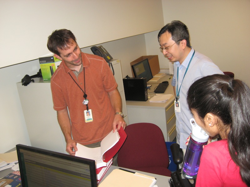 |
| Jacob Hooker, Changning Wang and Anisha Bhanot finalizing the eIND application to the FDA to request permission to use the probe in humans. Each copy was 1000 pages long! |
 |
| Changning Wang and Frederick "Al" Schroeder, the first authors of the initial two papers describing the probe and its potential applications. |