

The Daily Free Press recounts the HUBWeek event in which Center Director Bruce Rosen and medical illustrator Danny Quirk spoke about the intersectionality of human anatomy and visual art.
ISMRM Names Martinos investigators Junior Fellows Of The Society
August 6, 2015
Two young researchers — Christin Sander and Berkin Bilgic — were recognized for their already considerable contributions to science when they were named Junior Fellows by the International Society for Magnetic Resonance in Medicine. They received the honor at the society's 23rd annual meeting, held this June in Toronto.
The areas in which the two have made an impact reflect the diversity of work that is ongoing in the MGH Martinos Center for Biomedical Imaging.
 |
|
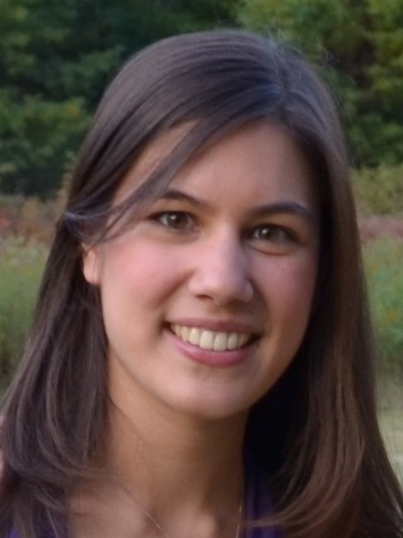
Christin Sander is developing and applying simultaneous PET and MRI technology.
|
Sander, a postdoctoral fellow working with Center Director Bruce Rosen, was recognized for her work with simultaneous PET and MRI, advancing the technology and applying it to answer a range of important neuroscience questions. Individually, these two imaging modalities offer complementary information: PET can image highly specific molecular events — how a drug binds to a specific receptor in the brain, for example. MRI and functional MRI can provide detailed anatomical information as well as the ability to track brain function through measures of hemodynamics. But combining them in a single scanner, with truly simultaneous acquisition, offers tremendous advantages over wielding each on its own — and in fact has opened up whole new fields of research.
Exploring this potential has been an important part of Sanders’ career thus far. She joined the Martinos Center as a graduate student in 2010, shortly after the installation of the Center’s first combined PET/MR scanner — at the time, one of only two in the United States (the Center now has two of its own). This timing enabled her to evolve with the fledgling PET/MR program there, beginning with an RF coil project working with hands-on hardware and eventually applying the technology to peer deep inside the living brain.
“Having an engineering background, I was immediately excited by the novel technological aspects of the integration of the scanners,” she said. “More recently, I have become fascinated about the variety of new applications this technology can have,” ranging from basic science applications such as exploring connections between receptor dynamics to brain function to elucidating mechanisms underlying psychiatric disorders.
Sander and colleagues continued to probe the inner workings of the brain, and in 2013 published a broadly influential paper in Proceedings of the National Academy of Sciences (PNAS). Among its significant findings, the study — the first to apply simultaneous PET/fMRI to investigate receptor dynamics — revealed correlations between receptor occupancy and measures of cerebral blood volume in space, time and dose in the D2/D3 dopamine system, which is implicated in reward and risk-taking behaviors.
The study could have important ramifications, she said. Not least: The use of in vivo surrogate markers of drug efficacy and dopamine (or other neurotransmitter) activity could lead to the development of new biomarkers for clinical use.
Working at the forefront of applying a new technology like this has proved tremendously rewarding for Sander. “I feel inspired thinking that our research facilitates an understanding of how the brain functions — how molecular changes lead to overall cognition — while pursuing the vision to find new treatment paths for diseases of the brain,” she said.
She completed her PhD last year and, as a postdoctoral fellow, has been extending her research exploring receptor dynamics with simultaneous PET/MRI. As described in abstracts submitted to the ISMRM meeting and to the recent BRAIN meeting in Vancouver, she and colleagues have looked at classification of in vivo drug function — to aid in determining drug efficacy — and the assessment of receptor desensitization and internalization. She presented the latter at the BRAIN meeting as a Niels Lassen Award finalist.
This work is only the tip of the proverbial iceberg for her. Sander and her collaborators are also pursuing further understandings of the mechanisms underlying the coupling of functional and hemodynamic signals in the dopamine and other receptor systems. At the same time, she is preparing to launch a study of the neurochemical signature and functional networks of deep brain stimulation with simultaneous PET/fMRI, funded by a recently received competitive Fund for Medical Discovery Postdoctoral Fellowship Award at MGH. And to all of this she is keen to add another angle — studying the effects of genetics on receptor dynamics and brain function — which promises to provide fresh new insights for a range of neurologic and psychiatric disorders.
 |
|
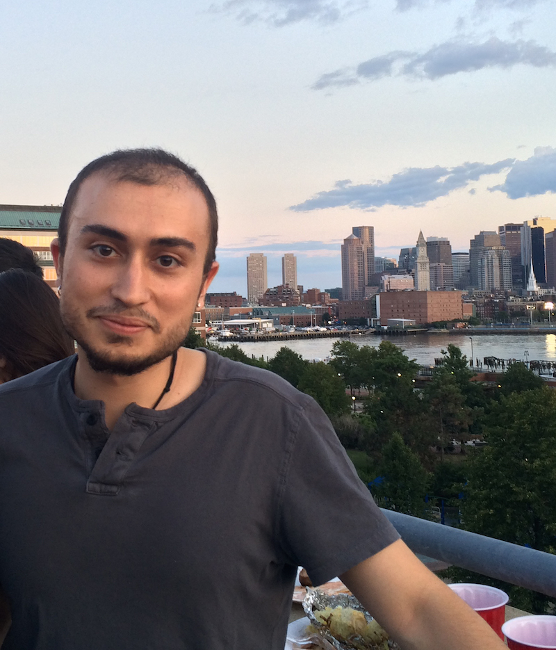
Berkin Bilgic is advancing methods that facilitate significant increases in the efficiency of MR imaging.
|
Berkin Bilgic, a postdoctoral fellow in the Center working with Kawin Setsompop, was recognized for his research developing data acquisition and reconstruction methods that promise dramatic increases in the efficiency of MRI. These methods for efficient 3-dimensional (3D) and simultaneous multislice (SMS) imaging will allow us to delve even deeper into the brain by providing much finer detail than was previously possible.
Any number of applications could benefit from these. In fact, the techniques are already providing better tools for scientists working in a variety of disciplines. And with further development, they could transform the acquisition of MR images in the clinic, leading to improved diagnosis, for instance, in epilepsy, stroke, multiple sclerosis and tumor imaging.
But what exactly are they, and how do they work?
Simultaneous multislice imaging refers to the imaging of more than one plane, or “slice,” at a time — as opposed to the single-plane imaging of conventional MRI. A few years ago, Setsompop and colleagues introduced the SMS technique Blipped-CAIPI, which gives investigators a means to acquire up to ten slices of brain scans at a time, allowing them to obtain much faster snapshots of what is going on in the brain. Now, Bilgic is working with Setsompop and his team to extend these capabilities with a technique called Wave-CAIPI for SMS as well as for 3D imaging.
Wave-CAIPI offers a greater than 10-fold increase in acquisition speed, Bilgic said, while substantially mitigating the noise amplification stemming from the reconstruction process of sub-sampled data. This striking improvement easily expands the horizons of what is possible for researchers working with MRI — it will enable whole-brain imaging with high resolution and high contrast, for example. Ultimately, it also holds the key to improved clinical exams.
Bilgic presented his recent progress with the technique at the ISMRM meeting in Montreal. The abstracts “RARE/Turbo Spin Echo Imaging with Simultaneous MultiSlice Wave-CAIPI” and “Rapid Multi-Orientation Susceptibility Mapping with Wave-CAIPI” describe some of the advances made possible with its introduction. Both won Summa Cum Laude awards at the meeting (in addition to supporting his selection as a Junior Fellow of the society), pointing up the potential of the new MRI method.
Neither he nor his collaborators are in any way resting on their laurels, though. They continue to develop the technique in ways that will have far-reaching implications for both research and clinical applications. In fact, they are already exploring ideas that could enable a greater than 20-fold improvement in MR acquisition speed. This has the potential to enable clinical translation of encoding-intensive novel contrast mechanisms, such as susceptibility anisotropy. Susceptibility has shown tremendous sensitivity to iron and myelin, and could provide the means to monitor and detect neurodegenerative diseases such as multiple sclerosis.
The dramatic, 20-fold acceleration would also be critical in advancing the method relaxation parameter mapping — an encoding-intensive technique that yields quantitative tissue parameters rather than the conventional contrast-weighted images.
Bilgic is driven in his work by the knowledge that the efficient 3D and SMS imaging methods he and the others are developing “are likely to have a direct impact on how images are acquired in the clinic,” he said. “It is true that there is substantial work to be done to translate the novel sequences and reconstruction algorithms we have come up with. But we are motivated to take this last step. We develop these techniques to provide better tools to the clinicians for improved diagnosis and to the neuroscientists for more detailed study of the human brain.”
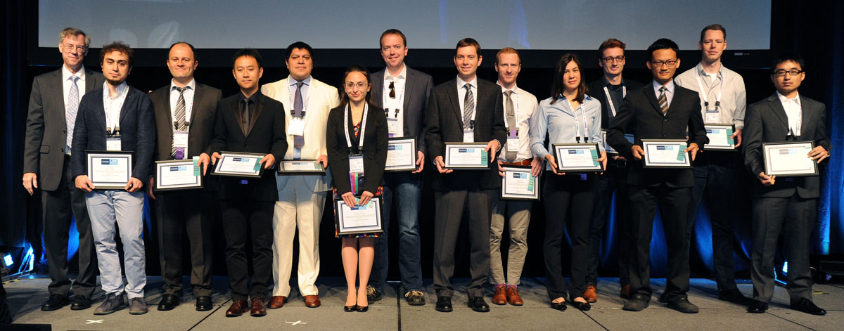
The Martinos Center's Berkin Bilgic (second from left) and Christin Sander (fifth from right) were named Junior Fellows of the International Society for Magnetic Resonance in Medicine (ISMRM)
-----
Subscribe to the Martinos Center newsletter to read more about what's happening in the Center.